
Research in the Au Lab
Cortical interneurons are specialized, locally projecting neurons that serve as the brain's primary source of inhibition—acting as the essential braking system of the cerebrum. Rather than sending signals across long distances, these cells work locally to regulate information flow. By controlling cortical rhythmicity and "gating" signal salience, they ensure the brain filters out background noise and processes important signals effectively.
The Au Lab investigates the complex development and synaptic integration of cortical interneurons. This intricate biological journey requires precise orchestration across three major developmental stages:
- Neuronal Lineage Specification: How stem cells decide to differentiate into specific types of interneurons.
- Long-Distance Cell Migration: How these newly born neurons travel from distant embryonic regions to their correct locations in the cortex.
- Synaptogenesis & Maturation: How interneurons form, refine, and maintain their synaptic connections throughout the cortex.
Why does this matter?
A growing body of evidence shows that early disruptions in interneuron development contribute directly to conditions such as autism spectrum disorder, schizophrenia, and epilepsy. Recent findings link the progressive loss of interneuron synapses to late-onset neurodegenerative disorders, including Alzheimer’s disease and frontotemporal dementia.
To capture the full picture of how interneurons integrate into brain circuits, our lab combines cutting-edge biological models with advanced computational tools.
Advanced Biological Models
Combining mouse genetics with human and mouse stem cell-based models (iPSCs) to capture key developmental mechanisms across species.
High-Resolution Imaging
Visualizing delicate neural structures using both in vivo (brain tissue) and in vitro (tissue culture) confocal microscopy.
Computer Vision & AI
Leveraging Synaptic Subtype Identification (SSID), our specialized analysis platform, to automatically categorize millions of synapses into distinct “synaptotypes” and map their spatial distribution.
Our current projects include
Stratification Across Subtypes
- Cortical interneurons are remarkably diverse, displaying a wide array of specialized roles and gene expression profiles. Our lab actively investigates how distinct interneuron subpopulations form synapses and utilize unique spatial strategies to "tile" the cortex during development. We hypothesize that disruptions in tiling and synapse-formation serve as major drivers in interneuron-related pathologies.
Synapses in Disease
- Interneuron dysfunction is implicated in a broad spectrum of neurological and psychiatric conditions, ranging from autism, schizophrenia, and depression to epilepsy and Alzheimer’s disease. To unpack these complex pathologies, we utilize Synaptic Subtype Identification (SSID) as a high-content quantitative readout of synaptic state. This enables us to pinpoint how interneuron synapses diverge between healthy and affected individuals, uncovering novel disease mechanisms and opening new avenues for therapeutic rescue. To ensure our findings translate directly to human health, we are actively expanding this approach to analyze interneuron synapses in human stem cell-derived models as well as postmortem human cortical tissue.
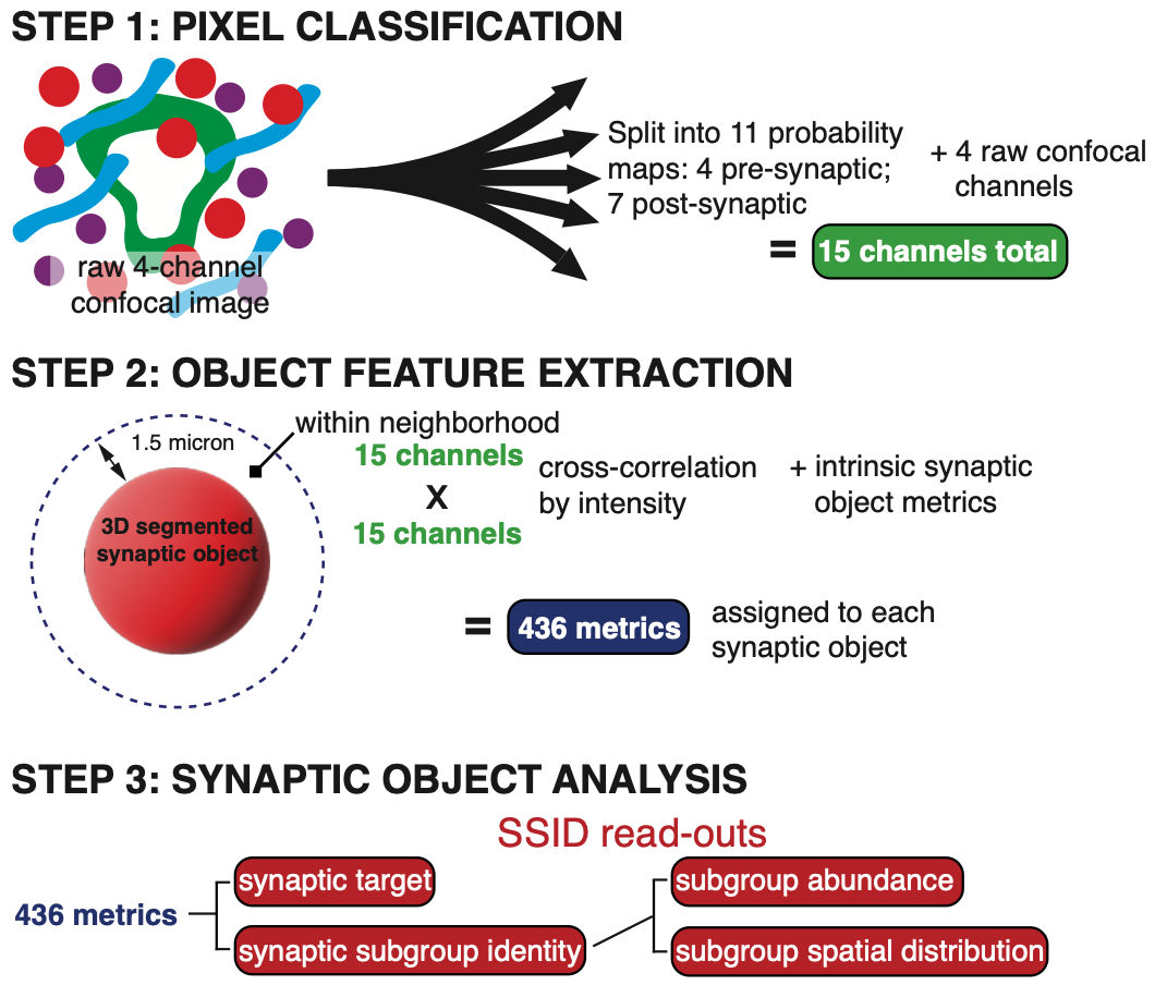
Overview of the SSID computer vision workflow for cortical interneuron synapse analysis (Figure above): 1. Feature Expansion: A 4-channel confocal image is processed through two machine learning models to generate 11 additional biologically relevant image channels. 2. Segmentation & Feature Extraction: Interneuron presynaptic boutons are segmented into discrete objects. Spatial and intensity-based relationships with their immediate microenvironment are evaluated, extracting hundreds of quantitative features per synapse to form a high-dimensional dataset. 3. Multi-tier Synaptic Analysis: Synapses are evaluated at both the individual level, classifying target specificity to identify distinct 'synaptotypes’; and the population level, assessing overall density, composition, and spatial distribution.